Femtosecond Laser-Assisted Cataract Surgery in Fuchs Endothelial Corneal Dystrophy: Long-term Outcomes
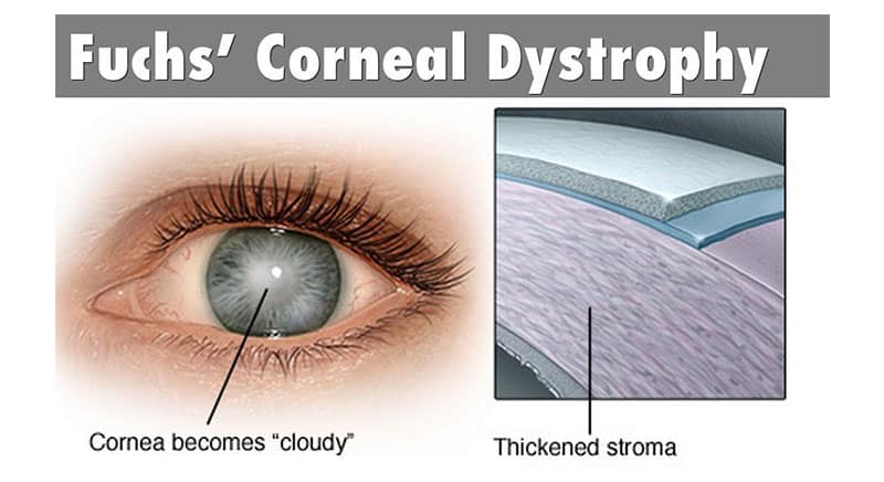
Fuchs endothelial corneal dystrophy (FECD) is characterized by progressive loss of corneal endothelial cells, thickening of Descemet’s membrane and deposition of extracellular matrix in the form of guttae. When the number of endothelial cells becomes critically low, the cornea swells and causes loss of vision. The clinical course of FECD usually spans 10–20 years. Corneal transplantation is currently the only modality used to restore vision. Over the last several decades genetic studies have detected several genes, as well as areas of chromosomal loci associated with the disease.
To our knowledge, no large-scale studies have evaluated the outcomes of femtosecond laser-assisted cataract surgery in patients with Fuchs endothelial corneal dystrophy.

PURPOSE:
To compare the corneal endothelial cell loss and central corneal thickness (CCT) after conventional phacoemulsification surgery or femtosecond laser-assisted cataract surgery in patients with Fuchs endothelial corneal dystrophy and senile cataract.
SETTING:
Xiamen Ophthalmic Center, Affiliate Xiamen University, Xiamen, China.
DESIGN:
Prospective case series.
METHODS:
Eyes with mild or moderate Fuchs endothelial corneal dystrophy and cataracts had femtosecond laser-assisted cataract surgery or phacoemulsification. The endothelial cell density (ECD), rate of ECD loss, cumulative dissipated energy (CDE), and CCT were measured preoperatively and 3 days and 1, 3, 6, and 12 months postoperatively.
RESULTS:
The study evaluated 31 eyes. The CDE was lower in the femtosecond group than in the phacoemulsification group (P < .05). The preoperative and postoperative ECDs were similar in the 2 groups (P > .05). The rate of ECD loss was higher in the phacoemulsification group from 1 to 12 months postoperatively (P > .05). The CCT was thicker in the phacoemulsification group 1, 3, and 6 months postoperatively (all P > .05). In both groups, the postoperative CCT at all follow-up visits were greater than the preoperative CCT (all P < .01). No bullous keratopathy or other intraoperative complications occurred in either group during the follow-up.
CONCLUSIONS:
For eyes with Fuchs endothelial corneal dystrophy and cataract, the CCT 12 months after surgery remained thicker than the preoperative thickness. The femtosecond group, with a lower CDE, tended to have a thinner CCT and less endothelial cell loss than the phacoemulsification group.
References: NCBI


